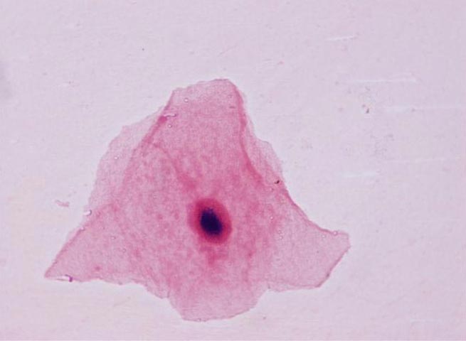
Squamous Epithelial cells (SECs)
At approximately 25-30 µm in size, SECs are the largest of the cellular elements that may be seen in a Sputum Q-score. They stain pale pink to red with the Gram stain. When SECs are seen in increased numbers in a Sputum Q-score it is indicative of a poor specimen (saliva). Squamous epithelial cells suggest the presence of superficial material and indicate the culture is likely to grow normal flora organisms of no clinical relevance. SECs main identifying characteristics are:
- Large, flat, "plate-like" cells
- Copious amounts of a clear or ground glass cytoplasm
- Relatively small dense nucleus, frequently located eccentrically
- Sharp and clear cut cellular boundaries with straight edges which give the cell a polygonal appearance
- Part of the cytoplasm may be folded back on itself
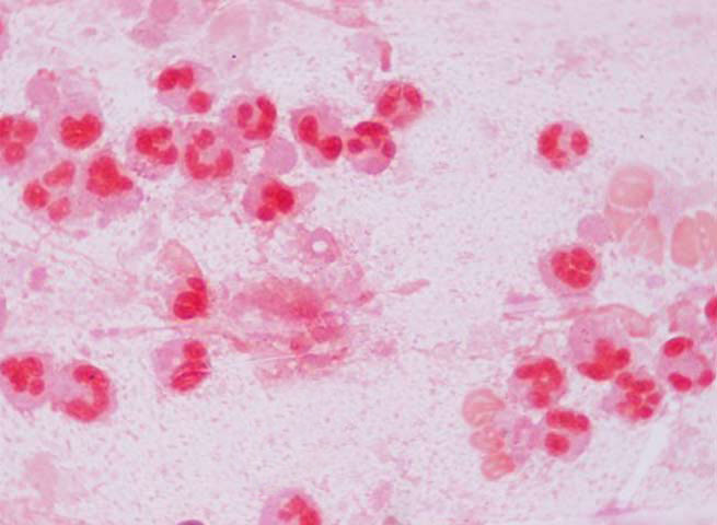
White blood cells (WBCs)
At 10-15 µm in size, WBCs are generally half to a third the size of SECs or smaller. They stain pink to red with the Gram stain. When related with bacterial infections, generally the type of WBC seen is a polymorphonuclear white blood cell (PMN). These PMNs have a multi-lobbed nucleus. The cytoplasm of PMNs is much smaller than the cytoplasm of SECs. It is generally approx. the same size as the nuclear material. Increased numbers of WBCs, specifically PMNs, are indicative of a good specimen and presence of an inflammatory process and possible infection.
PMN WBCs key identifying characteristics are:
- Multi-lobed nuclei
- Granular cytoplasm
- Cytoplasm sometimes contains phagocytosed bacteria
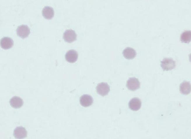
Red Blood Cells (RBCs)
RBCs are the smallest of the cellular elements demonstrated here. They are approximately 5-7 µm in size. They appear pale red on the Gram stain. They do not have a nucleus. They are not usually considered a significant finding in sputum Q-scores unless they are seen in elevated numbers (3-4+).
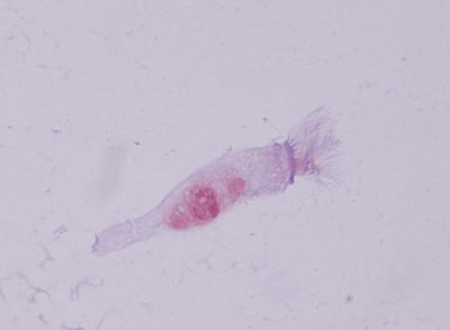
Respiratory Columnar Epithelial Cells
These cells are long and narrow compared to SECs. They occur in one or more layers. The cells are elongated and column-shaped. The nuclei are elongated and are usually located near the base of the cells. Some columnar epithelial cells are ciliated and possess fine hair-like outgrowths, i.e., cilia on their free surfaces. These cells are generally seen in specimens obtained mechanically from the bronchia and are therefore often indicative of a good quality specimen. Main identifying characteristics are:
- Long slender cells
- Cilia at one end of the cell
- Cilia supported by basal plate
- Nucleus located at base of cell
- Slender "tail" at opposite end to cilia
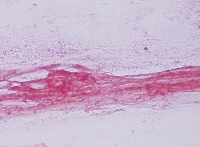
Mucous strands
Mucous strands are an important part of the normal defense mechanism of the lungs. The strands are produced by goblet cells which line the bronchi. They provide a transport system for the removal of foreign bodies from the respiratory tract. They spread in a thin, even layer over tissue surfaces.
In many disease states, an excess of mucus is produced and is expectorated. Cells and bacteria become enmeshed in the mucus and it is this combination of material that is known as sputum. Therefore the presence of mucous strands is indicative of a good quality specimen.